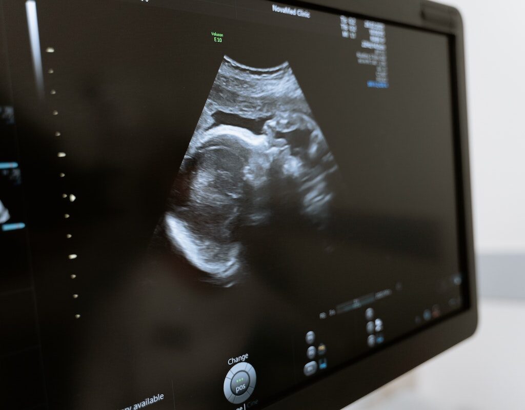
During pregnancy, you will have to go through different scanning and diagnostic tests to ensure your little one grows fine and healthy inside you. The TIFFA scan, which stands for Targeted Imaging for Fetal Anomalies (also known as Ultrasound Level II scan or the Fetal Anomaly Scan) is one of the essential scans conducted during pregnancy because it detects any congenital abnormalities in the growing fetus.
This anomaly scan helps visualize the presence of external and internal organs of the fetus. It allows your doctor to check if they are developing well.
Tiffa scan in which month of pregnancy?
This target scan is done during the second trimester which is between 18 and 23 weeks of pregnancy. It is an opportunity for you to see fetal movements; perhaps even catch a glimpse of your baby waving back at you!
The tiffa scan report shows detailed anatomy of the baby right from head to toe which includes:
Internal organs:
This detailed scan looks for the baby’s organs like the heart, brain, lungs, kidneys, stomach, and so on. This is done to check if the organs are formed well and functioning correctly. There are conditions like congenital heart disease, spina bifida (defect of the spinal cord), hernia, brain defects, congenital kidney anomalies like a baby with one kidney or without kidneys, bladder, gastroschisis – a condition in which the walls of the stomach do not form fully during pregnancy, etc. Your doctor would suggest further tests if any of these problems are detected during this early tiffa scan.
The external organs of the baby:
By the end of 18th week, your baby has begun rapid growth, and several of his or her external features would have formed well. During the TIFFA scan, your doctor would also collect images of your baby’s external organs and check for the development of organs such as eyelids, lips, fingers and toes, ears etc. This scan can detect common congenital problems like cleft lip and cleft palate. If these defects are present, your doctor will prepare you for the needed steps to manage the findings.
Position of your placenta:

The placenta is an organ that develops in your uterus during pregnancy. It acts as a link between mother and baby by providing oxygen and nutrients to your growing baby and removing waste products from your baby’s blood. Usually, the placenta attaches toward the top of the uterus, away from the cervix. However, problems can occur, such as the Placenta previa, which occurs when a baby’s placenta partially or covers the mother’s cervix — the outlet for the uterus. Placenta previa can cause bleeding throughout pregnancy and even during delivery. Hence it is essential to determine the position of the placenta to avoid complications and to have a smooth delivery.
Measure your baby:
The doctor will measure parts of your baby’s body during the scan to see how well he is growing. As a part of that, they will measure your baby’s head circumference (HC) and diameter (biparietal diameter or BPD) abdominal circumference (AC), femur or thigh bone (FL) and humerus or the arm bone. With these measurements taken, a doctor can easily match it with what’s expected for your baby at this stage of his development, depending on the due date. In this way, we can detect any deformities at the earliest and prepare for the necessary actions.

Check the level of Amniotic fluid:
Amniotic fluid is a clear, slightly yellowish liquid surrounding the unborn baby (fetus) during pregnancy. A sufficient amount of amniotic fluid is necessary as it covers the growing fetus in the womb and protects the fetus from injury and temperature changes. It also allows the freedom of fetal movement and musculoskeletal development. So, the Tiffa scan result will help doctors to understand the fetal condition better and provide treatment accordingly.
Chromosomal abnormalities:
A chromosomal abnormality occurs when a fetus has either the incorrect number of chromosomes, an incorrect amount of DNA within a chromosome, or structurally flawed chromosomes. These can result in congenital disorders like Down syndrome, Turner Syndrome, or a possible miscarriage. The anomaly scan helps to detect and pick up these chromosomal abnormalities in the unborn baby and gives your doctor a more precise picture in terms of the diagnosis.
Others:
During this anomaly scan, it is possible to determine the length of the birth canal, the position of the umbilical cord, and the blood flow to the womb. The tiffa scan price varies from city to city.
How and when is the Tiffa scan done?
The TIFFA scan is done during the second trimester. This is between 18 and 23 weeks of pregnancy.
There is no better feeling than the movement of life inside of you at this stage of pregnancy. A new baby is like the beginning of all things in your life: wonder, hope, and a dream of possibilities. This could be the start of more boundless happiness than we can imagine, but as with all good things in life, there will be some minor difficulties along the way.
Similar to the ultrasound scan, the preparations for the TIFFA scan consist of the following best practices.
- Wear comfortable and loose clothes
- Tiffa scan time: Around 45 minutes
- Drink lots of water so that the image is as clear as possible.
- Ultrasound works by means of sound waves. These waves require a medium to propagate and water serves as a good one. Because the pelvic organs are close to the urinary bladder, drinking water fills up the bladder so that it appears as a clearly separate solid structure. This makes it easy for clear visualization of the surrounding structures.
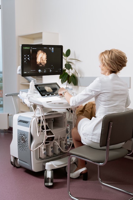
Since it is a scan of your abdomen, the technician will squeeze out a jelly and spread it on your tummy. The technician will then use a probe and slide it over your belly to pick up images of your womb and your baby.
These nine months of pregnancy prepare you both physically and mentally for the next phase of life and protect your baby, nurturing him or her inside your womb before you finally meet them. We hope you enjoy it!

